Medically reviewed by Dina Mohammed, MD · Last updated: October 7, 2026
Our Specialists for Moyamoya Disease and Syndrome
Moyamoya care at UChicago is delivered by a team that treats both adults and children, with decades of combined experience in direct and indirect bypass surgery. Experience matters here — bypass surgery for moyamoya is technically demanding, and outcomes are closely tied to surgeon volume.
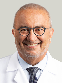
Dr. Awad directs the Neurovascular Surgery Program at UChicago and has spent four decades operating on complex cerebrovascular disease, including moyamoya. If you have moyamoya at UChicago, Dr. Awad is typically involved in planning the revascularization strategy and coordinating care across neurology, neuroradiology, and the operating room.
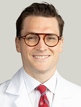
Dr. Polster performs direct, indirect, and combined bypass for adults and older children with moyamoya, and his NIH-funded lab studies the vascular biology of cerebrovascular disease. He works closely with Dr. Awad on the moyamoya service, handling both the microsurgical anastomosis and long-term follow-up imaging.
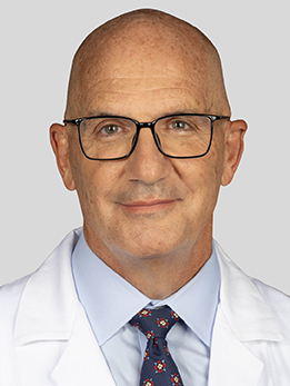
Dr. DiPatri is the pediatric neurosurgeon who sees children with moyamoya at UChicago Medicine Comer Children's Hospital, including kids with moyamoya syndrome from sickle cell disease, Down syndrome, and neurofibromatosis. He favors pial synangiosis and other indirect revascularization techniques that work particularly well in young, developing brains.
What Is Moyamoya?
Moyamoya is a rare cerebrovascular disease in which the large arteries that carry blood to the front of the brain — the terminal portions of the internal carotid arteries and their main branches — slowly narrow and eventually close off. In response, the brain grows a tangled network of tiny, fragile backup vessels to try to keep blood flowing.
On an angiogram, those collateral vessels look like a hazy cloud. The Japanese neurosurgeons who first described the disease in the 1960s called it moyamoya, which means 'puff of smoke' in Japanese. The name stuck.
When the narrowing happens on its own, with no underlying cause, we call it moyamoya disease. When it happens in someone who has another condition known to damage arteries — sickle cell disease, Down syndrome, neurofibromatosis type 1, prior radiation to the head, or certain genetic syndromes — we call it moyamoya syndrome. The surgical treatment is very similar, but the underlying disease matters for long-term management.
Moyamoya is most common in East Asia, but it occurs in every population. In the United States it shows up in two age peaks: children around age 5-10, and adults in their 30s and 40s. Women are affected slightly more often than men.
At a Glance
- Moyamoya is a progressive narrowing of the main arteries at the base of the brain, with fragile backup vessels forming a 'puff of smoke' pattern on angiography
- Children tend to present with mini-strokes or strokes from low blood flow; adults more often present with brain bleeding
- The best treatment is surgical bypass to restore blood flow — either direct (sewing a scalp artery to a brain artery) or indirect (laying vessels on the brain so new connections grow)
- A gene called RNF213 carries much of the risk in East Asian families, but moyamoya affects people of every background
- With experienced surgical revascularization, the risk of future stroke drops sharply and most patients return to normal activities
Seeking a review of a Moyamoya Disease and Syndrome diagnosis?
Your Moyamoya Disease and Syndrome imaging and clinical records can be directed to the appropriate specialist, often within 24 hours.
What Does It Feel Like?
Symptoms depend on age. Children and adults tend to present very differently, and recognizing the pattern is the first step toward getting the right test.
In children
Kids almost always present with symptoms of not enough blood flow to the brain — what doctors call ischemia. Parents often notice episodes that come and go:
- Transient weakness or numbness on one side of the body, sometimes triggered by crying, blowing out birthday candles, or hot soup (anything that causes rapid breathing and lowers blood CO2)
- Slurred speech or trouble finding words that lasts a few minutes and resolves
- Headaches, often severe and recurrent
- Seizures
- Involuntary movements (chorea) of an arm or leg
- A gradual drop in school performance or cognitive changes
- In young children, developmental delay or loss of previously learned skills
These spells are often mistaken for migraines, seizures, or behavioral problems for months before the diagnosis is made.
In adults
Adults more often present with sudden, dramatic events:
- Intracranial hemorrhage — bleeding into the brain or ventricles, causing sudden headache, confusion, weakness, or loss of consciousness. This is more common in adult moyamoya than in children.
- Ischemic stroke with sudden weakness, numbness, or speech problems that does not fully resolve
- Transient ischemic attacks (TIAs) — brief, stroke-like symptoms that recover
- Cognitive slowing, memory problems, or personality change from repeated small strokes
- Severe or new-pattern headaches
How Is It Diagnosed?
Most patients start with an MRI and MRA (magnetic resonance angiography) of the brain. MRI shows any strokes, old or new. MRA shows the blood vessels and can reveal the telltale narrowing of the carotid arteries and the wispy collateral network.
If moyamoya is suspected, a catheter cerebral angiogram is typically the next step. This test is considered the gold standard. A neurointerventional specialist guides a thin catheter to the brain's blood vessels and injects contrast material to produce a detailed, real-time map of all the arteries. This procedure enables your surgical team to clearly see which arteries are narrowed, how well the collateral network is developed, and what the target vessels on the brain's surface look like for a bypass.
We also evaluate the brain's ability to compensate. Perfusion imaging — using MRI, CT, or a specialized test like acetazolamide-challenge SPECT or BOLD-MRI — checks if the brain still has any'reserve.' If the brain has no reserve, it's at high risk for stroke and often requires surgery sooner rather than later.
Because moyamoya can run in families and because an RNF213 founder variant is strongly linked to the disease in East Asian populations, genetic testing and evaluation of first-degree relatives are increasingly part of the workup. In children, we also screen for the conditions associated with moyamoya syndrome — sickle cell disease, Down syndrome, neurofibromatosis, and others.
Types and Stages of Moyamoya
Disease vs. syndrome
- Moyamoya disease — the arterial narrowing occurs on its own, with no underlying cause. It is often bilateral and may have a genetic basis (RNF213 is the major susceptibility gene in East Asian populations).
- Moyamoya syndrome — the same vessel changes occur in someone with a predisposing condition such as sickle cell disease, Down syndrome, neurofibromatosis type 1, prior cranial radiation, Graves disease, or certain connective tissue disorders. Treatment of the underlying condition matters as much as the bypass.
Suzuki angiographic stages
In 1969 the Japanese neurosurgeons Suzuki and Takaku described six angiographic stages that trace the progression of moyamoya over time. Your surgical team will often refer to them:
- Stage I — narrowing of the internal carotid artery at its top, no visible collaterals yet
- Stage II — the characteristic 'puff of smoke' collateral network begins to appear at the base of the brain
- Stage III — collaterals become more prominent as the normal arteries narrow further; this is the stage at which many patients are first diagnosed
- Stage IV — the collaterals start to fade and new collaterals begin recruiting from the external carotid circulation
- Stage V — the classic moyamoya network further diminishes as external-to-internal collaterals take over
- Stage VI — the original moyamoya vessels are gone and the brain is perfused almost entirely through collaterals from the external carotid and posterior circulation
The stage helps predict how aggressive the disease is and how quickly you may need surgery, but it does not replace clinical judgment and perfusion imaging.
How Is It Treated?
Taking medication—like aspirin, controlling blood pressure, and managing risk factors—alone cannot halt the progression of moyamoya. For patients who have experienced symptoms, such as a stroke, TIA, or hemorrhage, and for many others with abnormal perfusion imaging, surgical revascularization is considered the standard treatment. The objective is to redirect blood flow from vessels outside the skull into the brain, thereby supporting the impaired circulation.
Direct bypass: STA-MCA
In a superficial temporal artery to middle cerebral artery (STA-MCA) bypass, the surgeon identifies a branch of the scalp artery you can feel pulsating in front of your ear, carefully dissects it free, and sews it directly into a small artery on the surface of the brain using sutures thinner than a human hair. The moment the clamp comes off, blood flows immediately through the new connection.
Direct bypass provides immediate improvement in blood flow. It is the preferred first option in most adults and works especially well when the target arteries on the brain are large enough to sew to.
Indirect bypass: EDAS, EMS, EDAMS, pial synangiosis
Indirect methods don't link directly. Instead, the surgeon places vascularized tissue—like a scalp artery, a muscle strip, or dura—right on the brain's surface, enabling new blood vessels to form within the brain over weeks to months. Typical techniques include:
- EDAS (encephaloduroarteriosynangiosis) — a scalp artery is laid onto the brain surface
- EMS (encephalomyosynangiosis) — a piece of temporalis muscle is laid onto the brain
- EDAMS (encephaloduroarteriomyosynangiosis) — a combination of artery, dura, and muscle
- Pial synangiosis — a refinement developed at Boston Children's, particularly well suited to children, in which the scalp artery is carefully sewn to the pia (the thin membrane covering the brain) to encourage ingrowth of new vessels
Indirect procedures are often the technique of choice in young children, whose vessels are too small for a reliable direct anastomosis and whose brains are especially good at growing new blood vessels.
Combined bypass
At UChicago and other centers, adults often undergo a combined direct + indirect bypass. This method provides the benefits of both techniques: immediate blood flow from the direct connection and enhanced long-term revascularization from the indirect part.
What the evidence says
For adult hemorrhagic moyamoya, the Japan Adult Moyamoya (JAM) Trial — the only randomized controlled trial in this field — showed that bilateral direct bypass reduced the rate of rebleeding and adverse events compared with medical therapy alone over 5 years of follow-up. For ischemic presentations and for children, large observational studies and meta-analyses consistently show that revascularization markedly lowers future stroke risk.
Medical management
Aspirin is commonly used to reduce the risk of small-vessel clots, particularly in ischemic moyamoya. Dehydration, low blood pressure, and hyperventilation should all be avoided because they further reduce blood flow to the brain. Blood pressure, cholesterol, and diabetes are managed aggressively.
Need another perspective on Moyamoya Disease and Syndrome?
Relevant specialists can review the Moyamoya Disease and Syndrome case together and provide one coordinated recommendation.
What Are the Outcomes?
Untreated, symptomatic moyamoya carries a high risk of repeat stroke or hemorrhage. After bypass surgery, that risk drops dramatically — but the procedure itself carries some perioperative risk, which is why experience matters.
| Outcome | With bypass | Without bypass | What to know |
|---|---|---|---|
| 5-year stroke rate (symptomatic adults) | ~5-10% | ~25-65% | Meta-analyses show a roughly 4-6x reduction in future stroke |
| 5-year rebleeding (adult hemorrhagic) | ~2.7% per year | ~7.6% per year | From the JAM Trial of bilateral direct bypass |
| Long-term stroke protection in children | >95% | Variable, high risk | Pial synangiosis series show durable stroke protection >20 years |
| Perioperative stroke (experienced centers) | ~3-5% | — | Most resolve; permanent deficits are less common |
Most patients feel and function better after revascularization — headaches improve, TIAs stop, and cognitive symptoms often stabilize or improve, particularly in children operated on early. The most important predictors of a good outcome are the severity of any baseline deficits, the quality of the operation, and how closely your long-term medical and imaging follow-up is managed. At UChicago, every moyamoya patient is followed with serial MRI and perfusion studies to ensure the bypass remains durable.
References
Have Questions About Moyamoya?
The team can review your Moyamoya Disease and Syndrome records, explain the available options, and help arrange the next step.
Schedule: (773) 702-2123