Medically reviewed by Irina Matache, MD · Last updated: October 9, 2026
Our Specialists for Pituitary Adenoma
Pituitary surgery is one of the most technically demanding procedures in neurosurgery — the tumor sits millimeters from the optic nerves, the carotid arteries, and the hormone-producing tissue you want to preserve. Outcomes at high-volume centers are meaningfully better, which is why our pituitary program pairs fellowship-trained skull base surgeons with dedicated neuroendocrinology, neuro-ophthalmology, and ENT partners.

Dr. Horowitz co-directs UChicago's Pituitary & Neuroendocrine Disorders Program and is fellowship-trained in skull base surgery at MD Anderson; for most patients who need pituitary surgery here, he is likely to be part of the operating team.
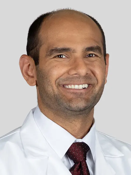
As Director of Endoscopic Neurosurgery, Dr. Ali specializes in the minimally invasive endonasal and keyhole approaches used for pituitary adenomas, with a practice focused on reaching deep skull-base lesions through narrow corridors that minimize disruption of normal tissue.

Dr. Yamini serves as Director of Neurosurgical Oncology and Vice Chair for Academic Affairs at UChicago, and co-manages pituitary tumor patients alongside his brain tumor and laboratory work on CNS drug delivery and tumor resistance mechanisms.
What Is a Pituitary Adenoma?
The pituitary is a pea-sized gland that sits in a small bony pocket (the sella turcica) right behind the bridge of your nose, directly below the optic nerves. Even though it's tiny, it controls much of your body's hormone system — thyroid, adrenal, reproductive, growth, and water balance. A pituitary adenoma is a benign tumor that grows from the hormone-producing cells of this gland.
Pituitary adenomas are among the most common brain tumors. On autopsy and imaging studies, small adenomas are found in about 10% of adults, but most of these are silent and never need treatment. The ones that do need treatment fall into two broad groups:
- Non-functioning adenomas — the tumor doesn't secrete active hormone, but as it grows it pushes on nearby structures (most importantly the optic chiasm) and crowds out normal pituitary tissue.
- Functioning (secretory) adenomas — the tumor over-produces one specific hormone, causing a recognizable clinical syndrome (for example, prolactinoma, acromegaly, or Cushing's disease).
Adenomas are also described by size: microadenomas are smaller than 10 mm, and macroadenomas are 10 mm or larger. Most non-functioning adenomas are only found once they've grown large enough to press on something — so by the time they come to attention, they're usually macroadenomas.
At a Glance
- Pituitary adenomas are benign tumors of the pituitary gland — the hormone "master gland" at the base of your brain
- They are common: imaging studies suggest roughly 1 in 10 adults has one, though most are small and never cause symptoms
- Some cause problems by pressing on nearby structures (especially the optic nerves), and others by overproducing a hormone
- Non-functioning adenomas typically need surgery; prolactinomas are usually treated first with medication, not an operation
- Endoscopic endonasal transsphenoidal surgery — reaching the tumor through the nose — is the standard approach when surgery is needed
Have recent studies for Pituitary Adenoma?
Call to arrange a specialist review of your Pituitary Adenoma imaging and records, often within 24 hours.
What Does It Feel Like?
Symptoms vary based on whether the tumor is causing issues through mass effect (compressing nearby structures), hormone excess, or hormone deficiency (impairing the normal gland). Many patients experience a mix of these effects.
Mass effect symptoms (most common in non-functioning adenomas)
- Vision loss, especially loss of peripheral vision on the outer sides of both eyes (called bitemporal hemianopsia) — you may notice bumping into things, or trouble seeing cars in the next lane
- Headaches, often dull and behind the eyes
- Double vision if the tumor reaches sideways into the cavernous sinus and presses on the nerves that move the eyes
- Sudden, severe headache with vision change or collapse — this can represent pituitary apoplexy (bleeding into the tumor), which is a neurosurgical emergency
Hormone deficiency symptoms
- Fatigue, cold intolerance, and weight gain (low thyroid)
- Low blood pressure, dizziness, nausea, or salt craving (low cortisol/adrenal)
- Loss of libido, irregular or absent periods, erectile dysfunction, infertility (low sex hormones)
- Excessive thirst and urination (diabetes insipidus — unusual before surgery, more common as a temporary post-op issue)
Hormone excess symptoms (functioning adenomas)
- Prolactinoma: milky nipple discharge, missed periods, infertility, loss of sex drive, erectile dysfunction
- Acromegaly (growth hormone excess): enlarging hands, feet, and jaw; rings and shoes no longer fitting; sweating; joint pain; sleep apnea; diabetes
- Cushing's disease (ACTH excess): central weight gain, round "moon" face, purple stretch marks, easy bruising, muscle weakness, high blood pressure, diabetes, mood changes
- TSH-secreting adenoma (rare): symptoms of hyperthyroidism — racing heart, weight loss, heat intolerance, anxiety
How Is It Diagnosed?
Pituitary adenomas are diagnosed with a combination of imaging, blood tests, and — when the tumor is near the optic nerves — formal visual field testing.
MRI with pituitary protocol
A dedicated pituitary MRI with and without contrast is the imaging test of choice. A standard brain MRI can miss small adenomas, so it's important that the scan uses thin slices through the sellar region and is interpreted by a neuroradiologist who reads a lot of pituitary imaging. The MRI tells us how big the tumor is, whether it's pushing up on the optic chiasm, whether it's invading the cavernous sinus, and how much normal gland is left.
Hormone testing
Every patient with a pituitary mass needs a full hormone workup. This is not optional — even "non-functioning" tumors need to have hormone secretion ruled out, because the treatment is completely different if the tumor turns out to be a prolactinoma. A typical panel includes prolactin, IGF-1 (for growth hormone excess), morning cortisol and ACTH, TSH and free T4, LH/FSH, testosterone or estradiol, and sometimes a 24-hour urine cortisol or overnight dexamethasone suppression test if Cushing's is suspected.
Visual field testing
If the tumor touches the optic chiasm, formal automated perimetry (a visual field test) by a neuro-ophthalmologist gives us an objective baseline. This matters for deciding whether surgery is urgent and for tracking recovery afterward.
Types of Pituitary Adenoma
Pituitary adenomas are grouped by which (if any) hormone they produce. The treatment, urgency, and outlook are very different from one subtype to the next — which is why hormone testing is critical before anyone operates.
Non-functioning adenoma (most common symptomatic type)
These tumors don't release enough active hormone to cause a clinical syndrome. They usually come to attention as macroadenomas pressing on the optic chiasm or crowding out the normal gland. Surgery is the first-line treatment when there's vision loss, significant mass effect, or progressive growth on imaging. Small, incidental non-functioning adenomas that aren't causing symptoms can often just be watched with periodic MRI.
Prolactinoma
The most common functioning adenoma. These tumors secrete prolactin and cause missed periods, infertility, low libido, erectile dysfunction, or milky breast discharge. Unlike almost every other brain tumor, prolactinomas are usually treated with medication, not surgery. Dopamine agonist pills (cabergoline, bromocriptine) shrink the tumor and normalize prolactin in the large majority of patients. Surgery is reserved for patients who can't tolerate the medication, don't respond to it, or present with sudden bleeding into the tumor (apoplexy).
Cushing's disease (ACTH-secreting adenoma)
An ACTH-secreting adenoma prompts the adrenal glands to overproduce cortisol, resulting in Cushing's disease. This condition is important to address, as it can lead to weight gain, diabetes, high blood pressure, weakened bones, a weakened immune system, and possibly early death if left untreated. Usually, these tumors are tiny microadenomas that can be hard to spot on an MRI, so specialized endocrine tests are vital for diagnosis. The main treatment method is transsphenoidal surgery to remove the adenoma.
Acromegaly (growth hormone–secreting adenoma)
GH-secreting adenomas cause acromegaly — gradual enlargement of the hands, feet, jaw, and facial features, along with joint pain, sleep apnea, hypertension, diabetes, and increased cardiovascular risk. The changes are slow enough that patients often don't notice them for years. Surgery is the first-line treatment, with medications (somatostatin analogs, GH-receptor blockers) used when surgery isn't curative.
TSH-secreting adenoma (rare)
These tumors produce thyroid-stimulating hormone and cause central hyperthyroidism. They're unusual (less than 1% of adenomas) and are also primarily treated with surgery.
How Is It Treated?
Endoscopic endonasal transsphenoidal surgery — the gold standard
When surgery is needed, the modern approach is the endoscopic endonasal transsphenoidal route. The surgical team — typically a neurosurgeon working alongside an ENT/skull base partner — goes through one or both nostrils, through the back wall of the sphenoid sinus, and directly into the sella. There's no incision on your face or head, no shaved hair, and no brain retraction.
Using a high-definition endoscope (a narrow, rigid camera) instead of a microscope gives a wide, panoramic, angled view of the tumor and the surrounding normal gland, carotid arteries, and optic nerves. For macroadenomas with suprasellar extension, the endoscope has been shown to improve the extent of tumor removal and visualization of tumor hidden behind bone or in corners.
Most patients go home within 2 to 3 days of surgery. You can usually return to light activity within a week or two, with restrictions on nose blowing, heavy lifting, and straining for about 4 to 6 weeks while the nasal reconstruction heals.
Medical treatment for prolactinoma
Prolactinomas are the exception to the "surgery first" rule. Dopamine agonist medications (cabergoline is usually first-line) normalize prolactin levels and shrink the tumor in the majority of patients, often dramatically. Most patients with prolactinomas never need surgery at all. Surgery is reserved for medication intolerance, resistance, apoplexy, or patient preference.
Radiation therapy
Radiation — usually delivered as stereotactic radiosurgery (a single highly focused dose) or fractionated radiotherapy — is used for tumor that can't be safely removed surgically, for recurrent tumor, or for functioning adenomas whose hormones remain uncontrolled after surgery and medication. It's effective at stopping tumor growth, but hormone remission can take years, and there's a gradual risk of developing new pituitary hormone deficiencies over time.
Endocrine management
Pituitary adenomas are as much an endocrine problem as a surgical one. Before, during, and after surgery, you'll work with a pituitary endocrinologist to identify any hormone deficiencies and replace them precisely — usually with hydrocortisone (for adrenal insufficiency), levothyroxine (for low thyroid), testosterone or estrogen (for low sex hormones), and occasionally desmopressin (for diabetes insipidus). Stress-dose steroids are given around the time of surgery for any patient whose cortisol axis might be at risk.
Watching and waiting
Not every pituitary adenoma needs treatment. If a small, non-functioning tumor is discovered by chance, doesn't press on any nearby structures, and isn't causing hormonal problems, it can often be tracked with routine MRI scans and lab tests. Treatment is only recommended if the tumor enlarges, causes vision or neurological symptoms, or starts affecting hormone levels.
Would a second review of your Pituitary Adenoma plan help?
The team can compare the available Pituitary Adenoma approaches and give you a coordinated recommendation.
What Are the Outcomes?
The outlook for pituitary adenoma is generally very good, especially when surgery is done by a high-volume team. Outcomes depend on the tumor's size, whether it has invaded the cavernous sinus, and the hormonal subtype. Here's what published series from experienced centers show:
| Tumor type | Gross total resection / remission | What to expect |
|---|---|---|
| Non-functioning microadenoma | ~80–90% | Excellent outcomes; often curative |
| Non-functioning macroadenoma | ~55–75% | GTR falls with cavernous sinus invasion |
| Prolactinoma (dopamine agonist) | ~80–90% biochemical control | Medication, not surgery, is first-line |
| Cushing's disease (microadenoma) | ~70–90% remission | Strongly surgeon-volume dependent |
| Acromegaly (microadenoma) | ~75–85% remission | Lower for macroadenomas; meds fill the gap |
Vision recovery
For patients who come in with vision loss from optic chiasm compression, roughly 80–95% experience improvement after tumor removal, and about a third recover completely normal visual fields. Younger patients, shorter duration of symptoms, and less severe pre-operative deficits predict the best recovery.
Complication rates
Endoscopic transsphenoidal surgery is generally very safe in experienced hands. Published rates from large modern series are approximately:
- Postoperative CSF leak: ~1–4% with modern closure techniques
- Permanent diabetes insipidus: ~1–3% (transient DI is more common but usually resolves within days to weeks)
- New permanent hypopituitarism: ~5–10%, depending on how much normal gland is preserved
- Meningitis: under 1%
- Major vascular injury or stroke: under 1%
- Operative mortality: well under 1% at experienced centers
These numbers vary meaningfully with surgical volume. Published data consistently show that high-volume pituitary surgeons have lower complication rates, higher remission rates, and shorter hospital stays than lower-volume centers — which is why where you have your surgery, and who does it, truly matters.
References
Have Questions About Pituitary Adenoma?
Questions about Pituitary Adenoma can be directed to the team for a focused review and next-step planning.
Schedule: (773) 702-2123