Medically reviewed by Irina Matache, MD · Last updated: October 9, 2026
Our Specialists for Ruptured Brain Aneurysm (Aneurysmal Subarachnoid Hemorrhage)
A ruptured aneurysm is one of the most time-sensitive problems in all of neurosurgery. At UChicago, a dedicated cerebrovascular team — trained in both open microsurgical clipping and endovascular coiling — is on call around the clock to secure the aneurysm and manage the days and weeks of complications that follow.
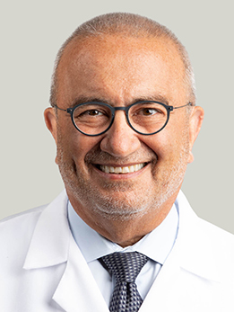
Dr. Awad is Section Chief of Vascular Neurosurgery at UChicago and directs the Neurovascular Surgery Program where ruptured aneurysm cases are managed around the clock. He served as senior investigator on the NIH-funded MISTIE III trial of minimally invasive surgery for hemorrhagic stroke, published in The Lancet (Lancet, 2019).
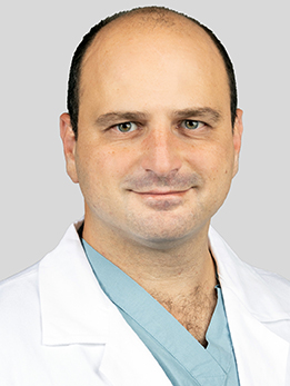
Dr. Doron is a dual-trained endovascular and open cerebrovascular neurosurgeon who brought more than 2,500 endovascular procedures from his Harvard/MGH fellowship to UChicago, and he coils or clips ruptured aneurysms depending on which approach is safer for the specific anatomy. He has published on intraoperative motor-evoked-potential monitoring to guide temporary clipping during open aneurysm surgery (Acta Neurochirurgica, 2023).
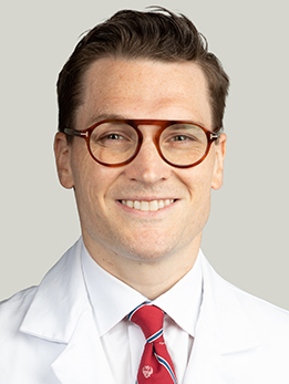
Dr. Polster is Co-Director of the UChicago Stroke Center and one of the cerebrovascular surgeons who manages ruptured aneurysm patients from the emergency department through the ICU course and rehabilitation. His NIH-funded laboratory studies the neurovascular unit and the gut-brain axis in hemorrhagic stroke.
What Is a Ruptured Brain Aneurysm?
A brain aneurysm is a weak, balloon-like bulge in the wall of an artery at the base of the brain. Most aneurysms cause no symptoms and are never found. But when one tears open, blood spills into the thin fluid-filled space surrounding the brain — the subarachnoid space. This is called an aneurysmal subarachnoid hemorrhage, or aSAH.
Aneurysmal SAH is rare but devastating. It affects roughly 6 to 9 people per 100,000 each year, strikes younger than most strokes (the average age is about 55), and carries a mortality of around 30-35% even with modern care. About 10-15% of patients die before ever reaching a hospital.
The good news: for the patients who make it to a comprehensive stroke center quickly, outcomes have improved dramatically over the last 20 years — driven by better imaging, endovascular coiling, and aggressive neurocritical care.
At a Glance
- A ruptured brain aneurysm causes a sudden, severe headache — often described as the worst of a person's life — and is a 911 emergency
- Diagnosis starts with a non-contrast CT scan of the head, sometimes followed by a spinal tap and a CT angiogram
- The aneurysm should be secured within 24 hours by either endovascular coiling or open surgical clipping
- Even after the aneurysm is fixed, the next two weeks are a marathon: watching for vasospasm, delayed stroke, and hydrocephalus in the ICU
- Outcomes depend heavily on how severe the bleed was at arrival and how experienced the team managing it is
Seeking a review of a Ruptured Brain Aneurysm (Aneurysmal Subarachnoid Hemorrhage) diagnosis?
Your Ruptured Brain Aneurysm (Aneurysmal Subarachnoid Hemorrhage) imaging and clinical records can be directed to the appropriate specialist, often within 24 hours.
What Does It Feel Like?
The hallmark symptom is a thunderclap headache — a sudden, severe, peak-in-seconds headache that is unlike any headache the person has had before. Patients often describe it as "the worst headache of my life" or "like being hit in the head with a bat."
Emergency warning signs
- Sudden, severe headache that reaches full intensity within a minute
- Nausea and vomiting
- A stiff neck, often developing a few hours after the headache starts
- Brief loss of consciousness or a seizure at the moment the aneurysm ruptured
- Sensitivity to light (photophobia)
- Blurred or double vision, or a drooping eyelid
- Weakness, numbness, or trouble speaking
- Confusion, agitation, or a change in behavior
The "sentinel headache"
In roughly 10-40% of patients, a smaller warning leak occurs days to weeks before the main rupture. This sentinel headache is often misdiagnosed as a migraine or tension headache. Any sudden severe headache that is different from a person's usual pattern deserves urgent evaluation — catching a sentinel bleed before a massive rupture can be lifesaving.
If you experience a sudden, severe headache, call 911. Do not drive yourself to the hospital.
How Is It Diagnosed?
Step 1: Non-contrast CT scan
The very first test is a non-contrast CT scan of the head. When performed within 6 hours of headache onset and read by an experienced radiologist, CT is essentially 100% sensitive for subarachnoid blood (BMJ, 2011). Fresh blood shows up bright white, filling the cisterns and sulci around the base of the brain.
Step 2: Lumbar puncture (if CT is negative)
If the CT is negative but clinical suspicion remains high — especially more than 6 hours after symptom onset — a lumbar puncture (spinal tap) is the next step. The doctor looks for red blood cells and a yellowish discoloration of the spinal fluid called xanthochromia, which confirms that blood has been in the cerebrospinal fluid for several hours.
Step 3: CT angiography to find the aneurysm
Upon confirming subarachnoid hemorrhage (SAH), a CT angiogram (CTA) — a CT scan with IV contrast — helps visualize the brain's blood vessels and pinpoint the aneurysm. Occasionally, a more detailed catheter angiogram (DSA) is necessary, involving the insertion of a thin tube from the groin or wrist to the brain's arteries. DSA is regarded as the gold standard and is usually the procedure during which endovascular coiling is performed.
Grading the severity
Two scales are used from the moment the patient arrives in the ED — they drive both prognosis and management:
- Hunt-Hess grade (I-V) — a clinical scale based on headache, stiff neck, level of consciousness, and weakness. Grade I is mild headache; Grade V is deep coma (J Neurosurg, 1968).
- Fisher grade (1-4) — a radiographic scale based on the amount and distribution of blood seen on the CT scan. It predicts the risk of vasospasm (Neurosurgery, 1980). A modified Fisher scale refined the prediction by accounting for intraventricular blood (Neurosurgery, 2006).
Hunt-Hess Grades at Presentation
When a patient arrives with an aneurysmal subarachnoid hemorrhage (SAH), the initial task for the medical team is to assign a Hunt-Hess grade. This evaluation is vital as it shapes the entire treatment strategy, determining the need for urgent angiography, the choice between coiling or clipping the aneurysm, the necessary intensity of ICU care, and the anticipated outcome.
Grade I
- What it looks like: Mild headache, slight neck stiffness, fully awake and oriented.
- What it means: The patient is neurologically intact. Excellent candidate for early aneurysm treatment; good prognosis.
Grade II
- What it looks like: Moderate to severe headache, stiff neck, possibly a cranial nerve palsy (for example, a drooping eyelid), but no weakness or confusion.
- What it means: Still a "good-grade" bleed. Most patients do very well with prompt treatment.
Grade III
- What it looks like: Drowsy or confused. May have mild weakness.
- What it means: Intermediate grade. Outcomes are still often favorable, especially if the aneurysm is secured quickly and the ICU course is smooth.
Grade IV
- What it looks like: Stupor, moderate to severe weakness on one side, early posturing.
- What it means: A "poor-grade" bleed. These patients are at high risk of hydrocephalus, vasospasm, and delayed stroke, and need aggressive ICU care. Recovery is possible but takes many months.
Grade V
- What it looks like: Deep coma, posturing, appearing moribund.
- What it means: The most severe grade. Some Grade V patients recover meaningfully — particularly after an emergency external ventricular drain and early aneurysm securing — but mortality remains high.
How Is It Treated?
Emergency stabilization
The patient is admitted to a neurological ICU. Blood pressure is carefully lowered to reduce the risk of re-rupture, while still keeping the brain perfused. If the patient is drowsy or has a large volume of blood, an external ventricular drain (EVD) is placed to relieve hydrocephalus and monitor intracranial pressure. Oral nimodipine, a calcium channel blocker, is started within 96 hours and continued for 21 days — it does not prevent vasospasm but it reduces the risk of delayed stroke and improves outcome (NEJM, 1983).
Securing the aneurysm — within 24 hours
Until the aneurysm is "secured," it can re-rupture at any moment, with catastrophic consequences. Current American Heart Association guidelines recommend that the aneurysm be treated as early as possible and ideally within 24 hours of presentation (Stroke, 2023). There are two ways to do this:
Endovascular coiling
A thin catheter is threaded through an artery in the groin or wrist all the way up to the brain. Soft platinum coils are packed into the aneurysm sac from inside, causing the blood to clot and sealing the aneurysm off from circulation. For many aneurysms — especially those in the back of the brain or in patients who are older or medically fragile — coiling carries a lower rate of disability than surgery. The landmark ISAT trial randomized 2,143 patients and found that those treated with coiling had a significantly better chance of being independent at 1 year (Lancet, 2002), a benefit that persisted at 18 years (Lancet, 2015).
Microsurgical clipping
In the operating room, a neurosurgeon performs a craniotomy, carefully exposes the aneurysm, and places a tiny titanium clip across its neck — permanently excluding it from the circulation. Clipping is the preferred approach for certain aneurysms: wide-necked middle cerebral artery aneurysms, aneurysms with a large associated hematoma that needs to be removed, and complex shapes that don't lend themselves to coiling. In the Barrow Ruptured Aneurysm Trial (BRAT), clipping achieved higher rates of complete aneurysm obliteration and lower rates of re-treatment than coiling, with comparable functional outcomes at 6 years for anterior circulation aneurysms (J Neurosurg, 2015).
At UChicago, the cerebrovascular team reviews every case together and recommends whichever approach is safer and more durable for that specific aneurysm. The decision is never "one size fits all."
The next two weeks: delayed cerebral ischemia and vasospasm
Once the aneurysm is secured, the focus shifts to the delayed complications of SAH. Starting around day 3 and peaking at days 7-10, the arteries at the base of the brain can constrict — a phenomenon called vasospasm — which can cause a new stroke. About 20-30% of patients develop delayed cerebral ischemia (DCI), a clinical deterioration from reduced brain blood flow that is the single biggest driver of long-term disability in SAH (Stroke, 2010). Management in the ICU includes:
- Daily neurological exams and transcranial Doppler ultrasound to watch for vasospasm
- Aggressive blood pressure augmentation (induced hypertension) when signs of DCI appear
- Endovascular rescue — intra-arterial verapamil or balloon angioplasty — for severe, medication-refractory vasospasm
- Management of hydrocephalus, often requiring a temporary or permanent CSF shunt
Rehabilitation
Recovery from SAH is a long road. Most patients spend 2-3 weeks in the ICU, followed by a stay on the neurosurgical floor, then weeks to months of inpatient and outpatient rehabilitation. Fatigue, memory problems, mood changes, and headaches can linger for a year or more — even in patients whose imaging looks fine.
Need another perspective on Ruptured Brain Aneurysm (Aneurysmal Subarachnoid Hemorrhage)?
Relevant specialists can review the Ruptured Brain Aneurysm (Aneurysmal Subarachnoid Hemorrhage) case together and provide one coordinated recommendation.
What Are the Outcomes?
Outcomes after aneurysmal SAH depend on three main factors: the severity of the bleed at presentation (Hunt-Hess grade), how quickly the aneurysm is secured, and how well the ICU team manages delayed complications. Here's what the data looks like when SAH patients are treated at high-volume comprehensive stroke centers:
| Hunt-Hess Grade at Arrival | 30-Day Mortality | Good Outcome at 6 mo (mRS 0-2) | What to Know |
|---|---|---|---|
| Grade I | ~2-5% | ~85-90% | Excellent; most return to normal life |
| Grade II | ~5-10% | ~75-85% | Good outlook with prompt treatment |
| Grade III | ~15-20% | ~55-65% | Intermediate; ICU course matters |
| Grade IV | ~30-40% | ~30-40% | Poor grade; aggressive care required |
| Grade V | ~50-70% | ~10-20% | Highest risk; recovery is possible but slow |
The modified Rankin Scale (mRS) is the standard way of measuring recovery after stroke. A score of 0 means no symptoms, 2 means independent in daily activities, and 6 means death. Most SAH outcome studies report the proportion of patients with a "good outcome" — mRS 0-2 — at 6 months.
Two things are important to understand about these numbers. First, they are averages across many centers, and outcomes at experienced comprehensive stroke centers are consistently better than the overall average. Second, there is enormous individual variation — Grade IV and V patients do sometimes walk out of the hospital, and the first 24-48 hours of care often make the difference. That's why where you are treated, and how quickly the team moves, matters so much.
References
Have Questions About a Ruptured Brain Aneurysm?
The team can review your Ruptured Brain Aneurysm (Aneurysmal Subarachnoid Hemorrhage) records, explain the available options, and help arrange the next step.
Schedule: (773) 702-2123